Human Respiratory System
Human Respiratory System
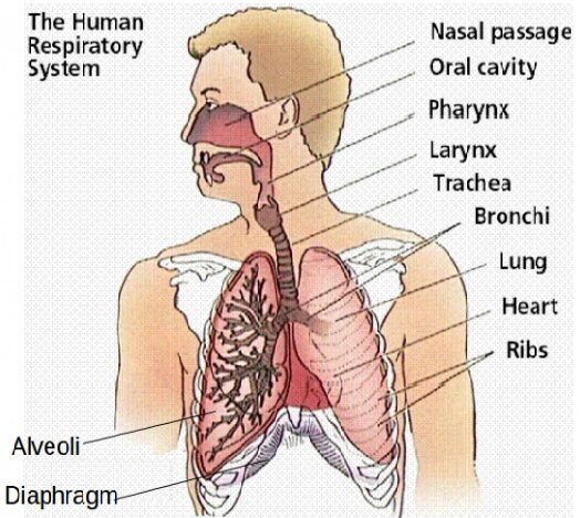
The human respiratory system is a highly specialized and intricate network of organs and structures that ensures the body gets the oxygen it needs and expels carbon dioxide, which is a byproduct of metabolism. Let’s dive into each part of the respiratory system in more detail.
2. Nasal Cavity and Nose
The nose is the primary entry point for air into the respiratory system, but the nasal cavity (internal passage) plays several critical roles:
- Air Filtration: Tiny hairs called cilia and the sticky mucous membrane in the nose trap particles such as dust, pollen, and bacteria, preventing them from entering the respiratory tract.
- Humidification: As air passes through the nasal passages, it is moistened by the mucus, which is important for protecting the delicate tissues of the lungs.
- Warming Air: The blood vessels in the nose warm the incoming air to body temperature, which helps prevent irritation in the respiratory tract.
- Olfaction (Sense of Smell): The nose also contains olfactory receptors that are responsible for our sense of smell.
- The sinuses are air-filled cavities located in the skull that connect to the nasal passages. They help lighten the weight of the skull and also contribute to the resonance of the voice.
2. Pharynx (Throat)
- The pharynx is a muscular passageway that connects the nasal cavity and mouth to the larynx (for air) and the esophagus (for food). It is divided into three regions:
- Nasopharynx: The upper portion, connected to the nasal cavity, allows the passage of air from the nose to the larynx. It also has openings for the Eustachian tubes (which connect the pharynx to the middle ear).
- Oropharynx: The middle portion, which is behind the oral cavity (mouth). This part serves as a common passage for both air and food.
- Laryngopharynx: The bottom portion, which leads to the larynx (for air) and the esophagus (for food).
- During swallowing, the epiglottis (a flap of tissue) covers the airway to prevent food or liquid from entering the trachea.
3. Larynx (Voice Box)
The larynx is located at the top of the trachea and plays multiple roles:
- Voice Production: It houses the vocal cords (also called vocal folds), which vibrate to produce sound when air passes over them.
- Protection of the Airway: The larynx functions as a protective mechanism by ensuring that food and liquids do not enter the trachea. It does so by triggering the cough reflex if any foreign particles enter the airway.
- Airway Control: The larynx helps in the regulation of airflow, ensuring that the air can move into the trachea and, eventually, the lungs.
- The epiglottis is a leaf-shaped flap of cartilage located at the entrance of the larynx. It prevents food and liquids from entering the trachea when swallowing.
4. Trachea (Windpipe)
- The trachea is a rigid, tube-like structure that extends from the larynx to the bronchi. Its walls are reinforced with C-shaped cartilage rings to prevent collapse. The trachea serves as the primary passage for air to move to the lungs.
- Mucous Membranes and Cilia: The inner lining of the trachea contains ciliated epithelium, which produces mucus. This traps dust, bacteria, and other particles, which are then moved upward to be swallowed or expelled by coughing.
- Smooth Muscle: The trachea contains smooth muscle that allows for some constriction and dilation, helping to regulate airflow.
5. Bronchi and Bronchioles
After the trachea, the airway branches into two main bronchi (left and right), which lead to the left and right lungs, respectively.
- Primary Bronchi: These are the first major branches that divide into smaller bronchi, with one bronchus leading to each lung. The right bronchus is shorter and more vertical, making it more likely to get foreign particles or food that accidentally enter the airway.
- Secondary and Tertiary Bronchi: As the bronchi continue to branch out into smaller and smaller passages, they become known as bronchioles.
- Bronchioles: These are smaller, thinner airways that do not contain cartilage but are surrounded by smooth muscle. They control airflow and can constrict or dilate.
The bronchioles end in tiny air sacs known as alveolar ducts, where gas exchange occurs.
6. Lungs
The lungs are the two primary organs of respiration. They are located in the thoracic cavity (chest) and are divided into lobes:
- Right Lung: Has three lobes (superior, middle, inferior).
- Left Lung: Has two lobes (superior, inferior) to accommodate the heart on the left side of the body.
Each lung is enclosed in a pleura (a double-layered membrane), which provides protection and reduces friction as the lungs expand and contract during breathing.
7. Alveoli (Air Sacs)
At the end of the bronchioles are alveoli, tiny, balloon-like sacs that are surrounded by a dense network of capillaries (small blood vessels). The walls of the alveoli are only one cell thick, allowing for efficient gas exchange between the air and the blood.
- Gas Exchange: Oxygen from the inhaled air diffuses across the alveolar membrane into the blood, while carbon dioxide diffuses from the blood into the alveoli to be exhaled.
- Surfactant: A lipoprotein substance called surfactant is produced by the alveolar cells. It reduces surface tension, preventing the alveoli from collapsing and making breathing easier.
8. Diaphragm
The diaphragm is a large, dome-shaped muscle that separates the chest cavity from the abdominal cavity. It plays a crucial role in breathing:
- Inhalation: During inhalation, the diaphragm contracts and flattens, increasing the volume of the thoracic cavity, which causes a decrease in pressure inside the lungs. This creates a vacuum that draws air into the lungs.
- Exhalation: During exhalation, the diaphragm relaxes and moves upward, reducing the volume of the thoracic cavity and pushing air out of the lungs.
Breathing Mechanism
Breathing, or pulmonary ventilation, involves two phases:
Inhalation (Inspiration):
- The diaphragm contracts and moves downward.
- The intercostal muscles (between the ribs) contract, causing the rib cage to expand.
- The lungs expand, creating a pressure drop in the thoracic cavity, causing air to flow into the lungs.
Exhalation (Expiration):
- The diaphragm relaxes and moves upward, while the intercostal muscles relax, causing the rib cage to shrink.
- This increases the pressure inside the lungs, forcing air out.
Gas Exchange Process (External Respiration)
Gas exchange occurs in the alveoli between the air and the bloodstream:
Oxygen: Oxygen diffuses from the alveoli into the blood, where it binds to hemoglobin in red blood cells and is carried to tissues and organs.
Carbon Dioxide: Carbon dioxide, a byproduct of metabolism, diffuses from the blood into the alveoli and is expelled from the body during exhalation.
Control of Breathing
- Breathing is controlled by the medulla oblongata and pons in the brainstem, which detect the levels of oxygen, carbon dioxide, and pH in the blood:
- When carbon dioxide levels rise or blood pH decreases (becoming more acidic), the brain signals the respiratory muscles to increase the rate and depth of breathing.
- Chemoreceptors in the aorta and carotid arteries also help monitor blood gases and send feedback to the brain.
Common Respiratory Disorders
Asthma: A chronic condition where the airways become inflamed and narrowed, making it difficult to breathe.
Chronic Obstructive Pulmonary Disease (COPD): Includes diseases like emphysema and chronic bronchitis, which cause chronic obstruction of airflow, often due to smoking.
Pneumonia: Infection that causes inflammation and fluid accumulation in the lungs, impairing gas exchange.
Lung Cancer: Malignant growths in the lungs, often linked to smoking and exposure to carcinogens.
Pulmonary Fibrosis: A condition in which the tissue in the lungs becomes scarred, leading to difficulty breathing.
The respiratory system is an incredibly complex network of structures that work together to ensure efficient gas exchange, regulate the body’s oxygen and carbon dioxide levels, and allow for the physical act of breathing. Without it, cells wouldn’t be able to generate the energy needed for the body to function properly.
Summary Chart — Respiratory System Structures & Functions
| Part | Location / Position | Structure / Key Features | Function |
| Nose / Nasal cavity | Front of head | Nostrils, nasal septum, conchae (turbinates), mucous lining, cilia | Filters, humidifies, and warms inhaled air; houses olfactory receptors |
| Sinuses | Skull bones around nasal cavity | Air‑filled cavities (frontal, maxillary, ethmoid, sphenoid) | Reduce skull weight, help warm/humidify air, voice resonance |
| Pharynx (Throat) | Behind nasal & oral cavities | Nasopharynx, oropharynx, laryngopharynx | Passage for air (and food in parts); connects nasal/oral cavities to larynx & esophagus |
| Larynx (Voice box) | At top of trachea | Cartilages (thyroid, cricoid, epiglottis), vocal cords | Produces speech, protects airway (epiglottis closes during swallowing) |
| Trachea (Windpipe) | From larynx into chest | C‑shaped cartilage rings, mucous membrane with cilia | Maintains open airway; filters particles; conducts air |
| Bronchi / Bronchial tree | Divisions inside lungs | Primary, secondary, tertiary bronchi → bronchioles | Distribute air to all lung regions |
| Bronchioles / Terminal bronchioles | Deep inside lungs | No cartilage, smooth muscle | Fine control of airflow to alveolar regions |
| Alveoli (Air sacs) | At end of bronchioles | Very thin walls, surrounded by capillaries | Site of gas exchange: O₂ in, CO₂ out |
| Lungs & Lobes | In thoracic (chest) cavity | Right lung (3 lobes), Left lung (2 lobes); pleura membranes | Houses bronchial tree and alveoli; enables gas exchange |
| Pleura & Pleural cavity | Surrounding lungs | Visceral + parietal pleura, pleural fluid | Reduce friction during breathing; keep lungs expanded |
| Diaphragm | Floor of thoracic cavity | Dome‑shaped muscle | Main muscle of breathing: contraction expands chest cavity |
| Intercostal Muscles | Between ribs | External and internal intercostals | Assist in expanding and contracting rib cage |
You can also read:
- Nutrition in organisms
- Reproduction in human
- Endocrine glands
- Common human diseases in humans due to deficiency of vitamins
Thank you for reading!!!


0 Comments