Human Digestive System
Human Digestive System
The human body is equipped with a complex digestive system designed to process food efficiently and extract essential nutrients necessary for health. This system comprises the alimentary canal and associated digestive glands, working in harmony to break down food and absorb nutrients.
Structure and Function of the Digestive System
- The digestive process begins in the mouth, where food is mechanically broken down by chewing and chemically by enzymes in saliva.
- The food then travels down the esophagus into the stomach, where gastric juices further digest it.
- In the small intestine, the majority of digestion and nutrient absorption occurs, aided by enzymes from the pancreas and bile from the liver.
- The remaining waste moves into the large intestine, where water is absorbed, and the remaining material is prepared for elimination.
- The digestive system of humans consists of an alimentary canal, which is associated with digestive glands.
Alimentary Canal
The human alimentary canal is a continuous muscular digestive tube measuring 8 to 10 meters that runs through the body.
- It opens at two ends with openings, which are the mouth at the anterior and the anus at the posterior end. It is highly coiled in certain regions.
- The alimentary canal consists of different parts such as the oral cavity, esophagus (food pipe), stomach, small intestine, and large intestine; specialized to perform different functions.
- Glands associated with the digestive system are salivary glands, liver, and pancreas.

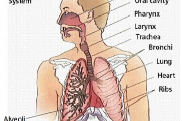
Labeled diagram of the alimentary canal of Human
Organs associated with the human digestive system
1. Oral Cavity
The oral cavity (buccal cavity) is the foremost part of the alimentary canal which opens to the outside by mouth. The mouth is bound externally by lips and cheeks, while internally by jaws bearing teeth. It helps in the ingestion of food and initiates digestion.
The cavity is associated with organs like teeth, tongue, and salivary glands; Minute quantities of water, water-soluble vitamins, and simple sugars like glucose (as in honey) are absorbed in the mouth. Saliva contains only a single enzyme Amylase (old name Ptyalin) which acts on starch.
- Teeth: An adult human has 32 permanent teeth, which are of four types: incisor, canine, premolar, and molar. The arrangement of teeth in each half of the upper and lower jaw in the order I, C, PM, and M is 2123/2123.
- Tongue: Tongue is a musculo-sensory organ present on the floor of the oral cavity. The surface of the tongue consists of several taste buds that can detect four types of tastes: salty, sour, sweet, and bitter. It also helps in ushing and swallowing or deglutition of moistened, partially digested food in the form of a small ball called a bolus.
2. Pharynx (or throat):
The mouth cavity opens into a small funnel-shaped cavity called the pharynx. It is a common passage for inhaled air and swallowing food.
3. Oesophagus
A narrow muscular tube arising from the pharynx, continuing through the thorax and ending in the stomach, food moves in the stomach through the esophagus in the form of the bolus through the movement of peristalsis. These peristaltic movements occur all along the gut.
- It is approximately 10 inches long.
- In the esophagus, the movement of food occurs in a regulated wavy manner because of the muscular lining of the tube.
- The muscles contract and relax rhythmically and push the food downwards. These movements called peristalsis, occur throughout the alimentary canal.
4. Stomach:
The esophagus opens into a J-shaped, thick-walled muscular sac called the stomach. It is a large organ present on the left side of the abdomen and expands on receiving the food.
- An elastic bag-like structure with highly muscular walls, located below the diaphragm. The muscular walls of the stomach help in mixing the food thoroughly with more digestive juices.
- It serves as the storehouse of food and a place where proteins are partially digested.
- The wall of the stomach contains many tubular glands termed gastric glands. These release the gastric juice into the lumen of the stomach which contains three types of secretions:
- Enzyme: A protein-digesting enzyme called pepsin breaks down proteins into smaller components, peptones in an acidic medium.
- Hydrochloric acid: It creates an acidic medium for pepsin to act and also kills harmful germs that may enter the stomach along with the food.
- Mucus: A slimy substance that protects the inner lining of the stomach from the action of the acid.
- The stomach churns the food mixing it with gastric juice and thus produces a creamy chyme (partially digested food). It contains Water (98%), some salts, hydrochloric acid (0.5%), lubricant mucin, and two enzymes pepsin. Water, glucose, ethanol (alcohol), certain minerals, vitamins, and certain drugs may be absorbed into the cells lining the stomach.
- This absorption occurs by osmosis, diffusion (down the concentration gradient), and active transport (against a concentration gradient
5. Small intestine:
The small intestine is a highly coiled narrow tube, the site of complete digestion and absorption of food.
- It is the longest part of the alimentary canal, a tube about 7 meters long and about 2.5 cm wide.
- The exit of food from the stomach is regulated by a sphincter muscle which releases it in small amounts into the small intestine. From the stomach, the food now enters the small intestine.
- The small intestine is the site of the complete digestion of carbohydrates, proteins, and fats. The liver and pancreas help the small intestine with digestion by their juices, The pancreatic enzyme makes the food alkaline, as it is acidic from the stomach.
- Bile juice comes from the liver which acts on fats as, with the help of bile, fat molecules break into small globules, and enzymes work on it effectively.
- pancreatic juice contains enzymes like trypsin for digesting proteins and lipase for breaking down emulsified fats. The enzymes present in it finally convert the proteins to amino acids, complex carbohydrates into glucose, and fats into fatty acids and glycerol.
- The inner wall of the small intestine has numerous minute folds (finger-like projection) called villi (singular, villus) further increasing the surface area of absorption,
- The epithelial cells have microvilli which are projections of the plasma membrane to further increase the absorptive surface.
- It is narrow for the slow movement of nutrients allowing absorption.
- Nutrients absorbed into the blood are carried by veins into the liver, and the nutrients absorbed by the lacteals (small lymph vessels) enter the lymphatic system.
- Duodenum–Short upper part, next to the stomach
- Jejunum–Slightly longer part, about 2 meters long.
- Ileum–Longest, about 4 meters long, coiled and twisted.
6. Large Intestine:
The large intestine is about 1.5 meters long, It does not secret any digestive enzymes. It has three parts:
(i) Caecum–Small blind pouch found at the junction of the small and large intestines. A narrow worm-shaped tube (vermiform appendix) projects from the caecum.
(ii) Colon: A little over 1 meter long, it has three parts termed ascending, transverse, and descending limbs of the colon.
(iii) Rectum: Last part, about 15 cm. long. It has two parts, the rectum proper and the anal canal. The anus is the external opening surrounded by circular muscles (sphincters).
Most of the water present in the food is absorbed in the colon by diffusion. Unabsorbed food is sent here, some mineral ions are absorbed by the colon through active transport, rest material is removed from here through the anus.
Note- The vermiform appendix is a vestigial (functionless) organ in humans, but is large and functional in herbivorous mammals.
Alimentary Canal Associated Glands
1. Parotid glands- located in front of and below each ear, produce watery saliva rich in amylase
2. Submaxillary glands– close to the inner side of the lower jaw, produce water and mucus
3. Sublingual glands –below the tongue, produce water and mucus.
4. Liver–Liver is the largest gland, located on the upper right side of the abdomen below the diaphragm. It secretes bile.

structure of liver
5. Pancreas– Pancreas is a large, cream-colored gland located in the bend of the duodenum. Its digestive secretion (pancreatic juice) is poured into the duodenum by the pancreatic duct. The pancreatic juice contains:
- Trypsin: Breaks down larger molecules of proteins to peptones, peptides, and amino acids.
- Lipase: Acts on emulsified fats and converts them into fatty acids and glycerol.
- Pancreatic amylase: Breaks down carbohydrates into simpler sugars.

Pancreas
Some enzymes and their role in the human digestive system:

Some enzymes and their roles
The Role of Balanced Nutrition in Human Digestive System:
Balanced nutrition is crucial for maintaining overall health and well-being. A diet that includes a variety of nutrients—such as carbohydrates, proteins, fats, vitamins, and minerals—supports bodily functions and promotes growth and development. For instance, proteins are essential for tissue repair, carbohydrates provide energy, and fats support cell function
In summary, the human digestive system efficiently processes food to extract essential nutrients necessary for health. Maintaining a balanced diet ensures that the body receives the nutrients it needs to function optimally. By understanding the digestive system and the importance of nutrition, individuals can make informed choices to support their health and well-being.You can also read:
- Nutrition in organisms
- Reproduction in human
- Endocrine glands
- Common human diseases in humans due to deficiency of vitamins
Thank you

1 Comment
fabrika soba · September 8, 2023 at 8:20 pm
Great information shared.. really enjoyed reading this post thank you author for sharing this post .. appreciated