The Viruses- Classification, Structure and Diseases
The Viruses- Classification, Structure, and Diseases
Viruses are sub-microscopic, acellular, and non-cytoplasmic infectious agents. These replicates are only in the living cell of an organism. Viruses can infect every type of life form like animals, plants, microorganisms like bacteria, etc. and these are transmissible from diseased to healthy organisms.
Dmitri Ivanvosky discovered the virus (1892) in an extract of a tobacco plant that was infected with the Tobacco mosaic virus (TMV).
- The study of Viruses is known as virology (under microbiology)
- The term virus was coined by Pasteur (Latin Virus- Venom)
- The latest definition of a virus is given as “Viruses are infective nucleoproteins”.
General Characteristics of Viruses
- All Viruses are obligate parasites and they can multiply only within the host cell.
- They contain only a single nucleic acid either DNA or RNA.
- The viruses are host-specific.
- The viruses are found in almost every ecosystem on earth and these are the most numerous type of biological entities.
Non-living characteristics of viruses
- No protoplasm is found
- enzymes are also not present
- respiration does not take place
- They can be crystalized
- In a culture medium, they cannot grow.
Living character of viruses
- They possess nucleic acid by this they are capable of synthesizing protein.
- they multiply inside the living cell
- causing diseases.
on the basis of these characteristics, it can be said that viruses are transitional groups between living and non-living.
Classification of Viruses
Viruses can be classified on the basis of phenotype characters morphology, nucleic acid, mode of replication, the host organism, and type of disease caused by them. Formally the viruses are classified by the ICTV (International Committee on Taxonomy of Viruses) system. Mostly Baltimore classification system was used, through it viruses were placed in seven groups on the basis of the pattern of RNA synthesis.
Baltimore Classification (1971)
In the Baltimore classification system viruses are placed in seven groups on the basis of nucleic acid (DNA or RNA), single-stranded or double-stranded, sense, and the method of replication. The classification is named after the Nobel prize-winning biologist David Baltimore. The seven groups are the following:
- I: dsDNA viruses (e.g. Adenoviruses, Herpesviruses, Poxviruses)
- II: ssDNA viruses (+ strand or “sense”) DNA (e.g. Parvoviruses)
- III: dsRNA viruses (e.g. Reoviruses)
- IV: (+)ssRNA viruses (+ strand or sense) RNA (e.g. Coronaviruses, Picornaviruses, Togaviruses)
- V: (−)ssRNA viruses (− strand or antisense) RNA (e.g. Orthomyxoviruses, Rhabdoviruses)
- VI: ssRNA-RT viruses (+ strand or sense) RNA with DNA intermediate in life-cycle (e.g. Retroviruses)
- VII: dsDNA-RT viruses DNA with RNA intermediate in life-cycle (e.g. Hepadnaviruses)
DNA viruses: The viruses have DNA as a genome (exception- DNA reverse transcribing viruses).
- In this type viruses with Double-Stranded DNA- a group I (e.g. chickenpox and herpes) and single-stranded DNA –group II are found.
The RNA viruses: consist of the RNA genome.
- The viruses of group III with a double-stranded RNA genome. e,g, Rotavirus.
- The viruses contain positive-sense single-stranded RNA genomes (group IV virus). e.g. Hepatitis A virus, rhinoviruses, poliovirus, foot, and mouth virus.
- The group V viruses- negative-sense single-stranded RNA genome. e.g. Ebola, and Marburg viruses.
Reverse transcribing viruses: These viruses encode reverse transcriptase.
- Group VI viruses with single-stranded RNA viruses replicate through DNA intermediate. e.g. Retrovirus (HIV is a member of this gr.)
- The viruses of group VII with double-stranded DNA genomes replicate using reverse transcriptase. e.g. Hepatitis B virus.
Holmes’s classification of viruses
The Holmes Classification (1948): Holmes classified viruses into three groups as follows:
- Phaginae (the viruses that attack bacteria)
- Phytophaginae (attacks plants)
- Zoophaginae ( attacks animals)
This system was not accepted as morphological similarities were neglected in it.
The general structure of viruses
Viruses are inert outside the host cell. Small viruses, e.g., polio and tobacco mosaic virus, can even be crystallized. Viruses are unable to generate energy. As obligate intracellular parasites, during replication, they fully depend on the complicated biochemical machinery of eukaryotic or prokaryotic cells. The main purpose of a virus is to deliver its genome to the host cell to allow its expression (transcription and translation) by the host cell.
The infectious virus is called a virion. The simplest virions consist of two basic components:
- nucleic acid (single- or double-stranded RNA or DNA): either DNA or RNA
- Capsid – A protein coat, that functions as a shell to protect the viral genome from nucleases and which during infection attaches the virion to specific receptors exposed on the prospective host cell
- Capsids are formed as single or double protein (capsomeres) shells and consist of only one or a few structural protein species.
- Therefore, multiple protein copies must self-assemble to form the continuous three-dimensional capsid structure.
- The self-assembly of virus capsids follows two basic patterns one is helical symmetry, in which the protein subunits and the nucleic acid are arranged in a helix, and the second icosahedral symmetry, in which the protein subunits assemble into a symmetric shell that covers the nucleic acid-containing core.
Envelop: Some large virions possess glycoprotein envelopes surrounding the capsid called envelopes. It is made up of two lipid layers (lipoprotein bilayer) that may contain material of the host membrane or its own, it is not found in all viruses. The viruses are called enveloped e.g. influenza
Structure of TMV (Tobacco mosaic virus): (The bacterial virus)
TMV is a plant virus, a Tobacco mosaic virus. Ivanoski reported in 1892 that extracts from infected leaves were still infectious after filtration through a Chamberland filter candle.
- Beijerinck, in 1898, was the first to call ‘virus’, the incitant of the tobacco mosaic.
- He showed that the incitant was able to migrate in an agar gel, therefore being an infectious soluble agent or a ‘contagium vivum fluidum’ and definitively not a ‘contagium fixum’ as would be a bacterium

TMV virus
- It is rod-shaped, approx. 300nm in length and ~18 nm in diameter.
- Made up of RNA and Proteins.
- The RNA is single-stranded, helical, and coiled around the hollow axis of the rod.
- The capsid or protein coat is made up of 2130 molecules of the coat protein.
- The TMV is of naked type, i.e. envelope is not found, and the capsid is not surrounded by an envelope.
Structure of bacteriophage:
These viruses are intracellular parasites of bacteria, so-named on it, bacteriophages. In the 20th century, Fredrick Twort (an English bacteriologist) discovered viruses that infect bacteria. Later named bacteriophages.
Bacteriophage T4 is one of the seven Escherichia coliphages (T1–T7, T for type), which, in 1944, were suggested by Delbruck and coworkers to be models for study by the phage community
Bacteriophage T4 is classified as a member of the Myoviridae family of the Caudovirales order because it has a contractile tail.
- These may have DNA or RNA, but the shape of capsid varies as isohedral, filamentous, and head-tail-shaped.
- T- series bacteriophages (T3, T4, T6, etc.) are most studied, these are characterized by the presence of a tail.

Structure of Bacteriophage
- They consist polyhedral head, a short collar, and a helical structure tail.
- Head: it consists of about 2000 capsomeres with double-stranded DNA. The diameter of the head is 650 A.
- Tail: The 925 Å-long tail is surrounded by the contractile sheath and ends with a hexagonal base plate. Both tube and sheath are attached to the dome-shaped baseplate at the end away from the head. These tail fibers help bacteriophages for attaching to bacteria. These together form the baseplate that then nucleates the assembly of the tail tube around which the sheath assembles. The tail is entirely made up of proteins.
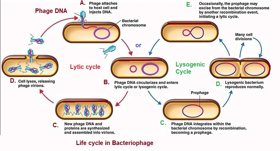
The life cycle of Bacteriophage
The life cycle of bacteriophages
from the initial stage of infection of bacteria to killing bacteria for releasing the virus’s progeny goes through certain stages given below:
- Adsorption: attachment of viruses’ tail fibers to a specific receptor on the surface of the bacterial cell.
- Penetration: The muramidase of phage works for the weakening of the cell wall and hollow-core penetrated through it, by the process of the DNA injected inside the bacterial cell.
- Multiplication or synthesis: The Phage DNA replicates and also synthesizes new proteins for its capsid. The subunits of the head, tail, and protein appear. some specific enzymes work in synthesis known as early proteins.
- Maturation and assembly of phage: The head and tail on maturation assemble, and phage DNA is surrounded by the protein coat. The formation of virion takes place by the addition of a tail.
- Lysis: The bacterial cell bursts by the process called lysis and new phage particles are liberated. These particles are able to infect other bacteria of the same type.
Some Viruses and Their Genetic Material
Some viruses with the type of nucleic acid (genetic material):
| Virus | Type of nucleic acid | Virus | Type of nucleic acid |
| Herpes | dsDNA | Measles | ssRNA |
| Chickenpox | dsDNA | Mumps | ssRNA |
| Hepatitis B | dsDNA | Polio | ssRNA |
| Cyanophages | dsDNA | TMV | ssRNA |
| Influenza | ssRNA | Mycophase | dsRNA |
| Rabies | ssRNA | Reovirus | dsRNA |
| HIV | ssRNA | Wound tumor virus | dsRNA |
Plant disease by Viruses
Some common plant diseases caused by viruses are given in the table below:
| Diseases | Causing agent |
| Tobacco mosaic | TMV |
| Papaya mosaic | Papaya mosaic virus |
| Cucumber mosaic | Cucumber mosaic virus |
| Potato mild mosaic | Potato virus X |
| Potato rugose mosaic | Potato virus Y |
| Potato leaf curl | Potato leaf curl virus |
| Potato leaf roll | Potato leafroll virus |
| Little leaf of brinjal | Brinjal little leaf virus |
| Rosette of groundnut | Groundnut mosaic virus |
| Sugar can mosaic | Sugarcan (sacchraum ) virus |
Animal diseases by Viruses
Some important animal viral diseases are given in the table:
| Diseases | Causal agent |
| Common cold | Rhinovirus |
| Influenza | Influenza virus |
| Rubella (measles) | Rubella virus |
| poliomyelitis | Poliovirus |
| Smallpox | Variola virus |
| Yellow fever | arbovirus |
| Chickenpox | Varicella virus |
| AIDS | HIV |
Common routes of viral infection
The common route through viral infection takes place in humans and the names of viruses are as follows:
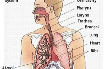
- Through respiratory route (i.e., droplet, aerosol, and respiratory secretions on the hands and elsewhere; oral exchange): influenza virus, varicella-zoster virus, human rhinovirus, human adenovirus, respiratory syncytial virus, parainfluenza virus, metapneumovirus
- By fecal or oral route: polioviruses, coxsackieviruses, hepatitis A virus, rotavirus, astrovirus, norovirus
- By direct contact with infected: human papillomavirus (HPV), molluscum contagiosum, herpes simplex virus type 1 (HSV-1)
- Through sexual transmission: human immunodeficiency virus type 1 (HIV-1), human T-lymphotropic virus type 1 (HTLV-1), hepatitis B virus (HBV), human papillomavirus types 16 and 18 (HPV-16, HPV18), HSV-2
- Urine-associated: cytomegalovirus (CMV)
- By parental route (i.e., blood and blood products, transplantation, tattooing, and scarification): HIV-1, HBV, hepatitis C virus (HCV)
- Bite of animals: rabies virus, Duvenhage virus
- Vertical route (e.g., germline, intrauterine, perinatal, human milk): HIV-1, HTLV-1, germline transmission of endogenous retroviruses
- Arthropod-borne route (e.g., mosquitos, ticks, sandflies): Japanese encephalitis virus, West Nile virus, dengue virus, yellow fever virus, Zika virus, chikungunya virus, and many others
- transmission associated with rodents: Lassa fever virus, sin Nombre, and other hantaviruses (e.g., Hantaan virus, Seoul virus, and Puumala virus)
- transmission associated with bats: rabies virus, Nipah virus, Ebola virus, severe acute respiratory syndrome coronavirus (SARS CoV)
- Monkey-associated transmission: herpes B virus, monkeypox virus, orf virus
- Other zoonotic associations (e.g., cows, sheep): orf virus, cowpox virus
You can also read:
- Common human diseases
- Nutrition
- Chemical classification of hormones
- Endocrine Gland And Hormones
- Common Human Diseases (pathogenic)
References: https://www.ncbi.nlm.nih.gov/books/NBK8174/
Thank you 🙂

4 Comments
Biological classification - PCSSTUDIES - Biology PCSSTUDIES · May 20, 2021 at 12:33 pm
[…] VIRUSES, VIROIDS, PRIONS, AND LICHENS […]
Zika Virus - PCSSTUDIES - Current Affaires Zika Virus · July 11, 2021 at 1:55 am
[…] The Viruses- classification, structure, and diseases […]
Common Human Diseases and Pathogenes - Biology · January 2, 2023 at 3:53 pm
[…] You can also read: Viruses […]
Common Human Diseases - Biology · May 19, 2023 at 9:32 pm
[…] Common human diseases caused by viruses […]